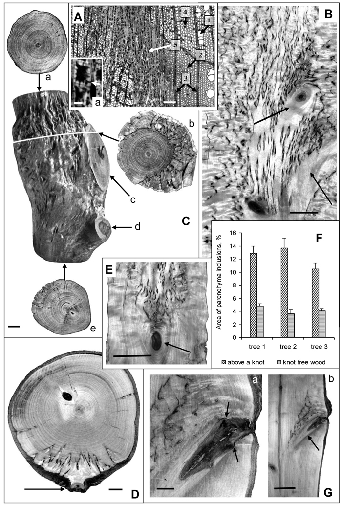
Fig. 1. Karelian birch wood at branch attachment sites.
А – microphotograph of a wood cross-section. 1 – group of three vessels, 2 – fibres, 3 – radial ray parenchyma, 4 – axial parenchyma, 5 – abnormal aggregation of parenchyma cells. a – a group of cells from the zone of parenchyma aggregation; arrow points to a pore in the walls of neighboring cells. Bar: A – 100 μm, a – 5 μm.
B – longitudinal section of a trunk. Arrows point to knots left of nearly perpendicular branches. The density of parenchyma inclusions above the knots is higher than in the rest of the trunk wood. Bar – 2.5 cm.
С – a debarked trunk segment of a 20-year-old tree: a – the wood at the top end of the trunk segment which displays only slight signs of figure; b – a transverse saw cut of wood in the zone of large branch attachment. The most valuable figure is situated on the side adjoining the branch. The darker portion of bark inside the wood indicates the border between branchwood and trunkwood; c – the wood of a large branch with no signs of figure, d – the figureless wood of a thin branch, e – the wood at the bottom end of the trunk segment with no figure below the zone of small brunch attachment to the trunk. Bar – 1 cm.
D – the surface of a trunk cross-cut of a 30-year-old Karelian birch tree. Parenchyma inclusions (figured wood) is found only in the narrow sector associated with the branch-trunk junction. The arrow points to the location of a thin branch attachment. Bar – 1 cm.
E – longitudinal trunk section. Parenchyma inclusions are confined to area above knot (indicated by arrow). Bar – 5 cm.
F – The area of parenchyma inclusions (%) in the middle part of the Karelian birch trunks (trees N 1–3). In each case knot-free figured wood zones were compared with figured wood above knots. M ± SE.
G – longitudinal trunk section. Arrows point to the site where the branch had been removed, causing the formation of parenchyma inclusions to stop. Bar: a – 1 cm, b – 5 cm.
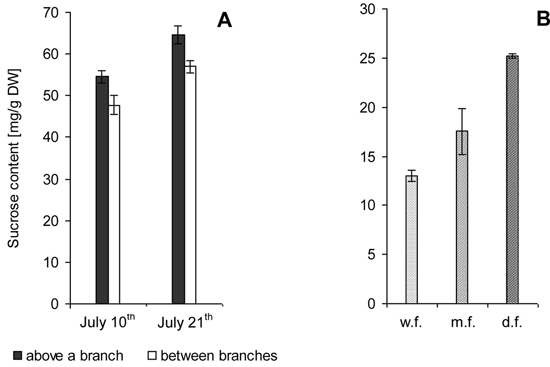
Fig. 2. Sucrose content in the zone of xylem growth and differentiation on the trunks of Karelian birch trees. M ± SE.
A – 7-year-old trees. Samples were taken from branch attachment sites and along the trunk between branches during active formation of wood structural abnormalities.
B – 22-year-old trees. Trunks with weakly figured wood (w.f.), medium figured wood (m.f.) and densely figured wood (d.f.).
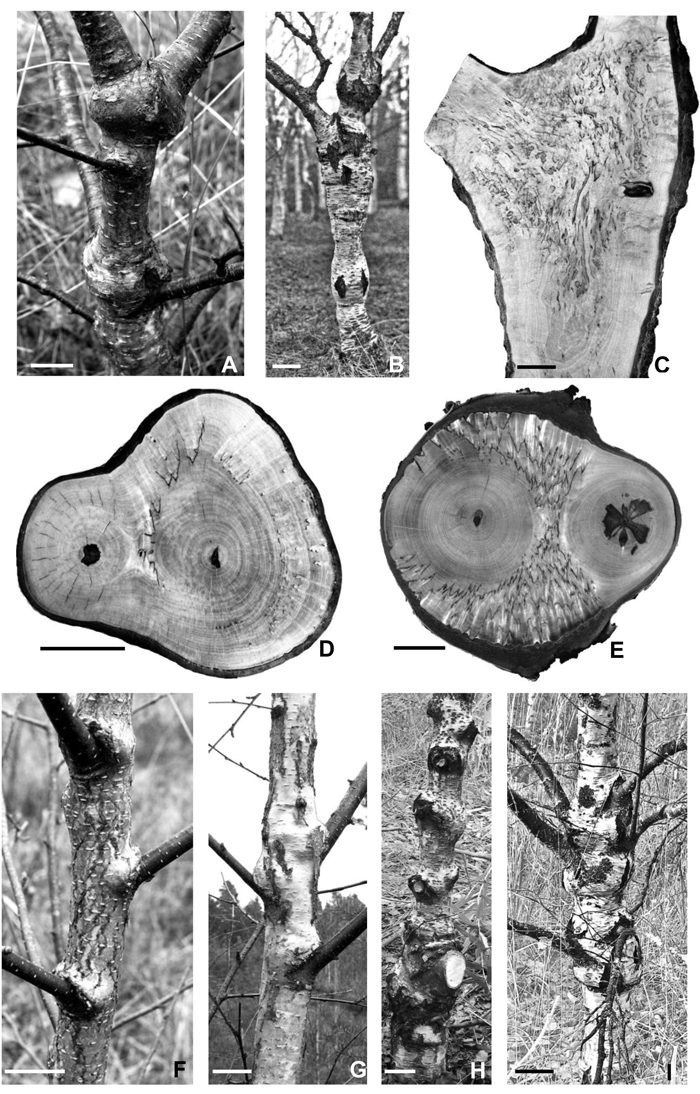
Fig. 3. Fork zones and different stages of globular bulges development on the trunks of Karelian birch trees.
A – a fragment of a 7-year-old stem with a well developed globular bulge located below the fork. Bar – 3 cm.
B – massive globular bulge below the fork in the trunk of a 35-year-old tree. Bar – 10 cm.
C – longitudinal section of a “figured” Karelian birch trunk at the branch junction site; sawn along the edge of a globular bulge, avoiding branchwood. Bar – 5 cm.
D, E – transverse saw cuts in the zone where two branches join. The wood of branches demonstrates no signs of figure. D – a few large parenchyma inclusions were found only in the trunk bifurcation zone. Bar – 5 cm. E – figured wood of the highest decorative value is situated in the branch attachment site. Bar – 3 cm.
F – the appearance of so-called “cheeks” over the sites of branch attachment detected on the trunk of a 5-year-old tree. Bar – 3 cm.
G – expansion of the swellings around the circumference of the trunk in a 10-year-old tree. Bar – 4 cm.
H – the formation of globular bulges at branch-trunk junctions on the trunk of a 17-year-old tree. Bar – 7 cm.
I – massive bulge formation in the zone of the attachment of a few large branches to the trunk of a 27-year-old tree. Bar – 10 cm.
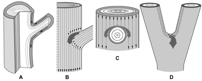
Fig. 4. Schematic representation of the confluence of sucrose phloem streams in the zones of branch attachment and the trunk fork. The xylem is white, the phloem is gray.
A – a fragment of a longitudinal section of the branch attachment area. Arrows indicate the pathways of phloem streams. On the lower side of branch attachment phloem flows move unobstructedly, whereas on the upper side of the branch-trunk junction there is an obstacle to phloem transport.
B, C – fragments of trunk surface with the zone of branch attachment. B – the scheme shows how the phloem flows of a branch and the trunk (thin dotted arrows) meet and merge into a common pathway (thick dark gray arrow). The resulting phloem pathway is reoriented to circumvent the branch. It provides for a rise in the sucrose level in tissues located sideways from the branch, inducing the formation of swellings. C – phloem flows of the trunk circumvent a branch.
D – a fragment of a trunk in the fork zone. Thin dotted arrows indicate the phloem flows of two axes which merge into a common pathway (thick dark gray arrow) at the top of the fork. It creates the conditions for a rise in sucrose content there, which induces abnormal tissue formation.